Featured post

Studies of East Atlantic bottom fauna

The University Museum of Bergen has acquired benthic samples from the West African continental shelf in agreement with the Guinea Current (GCLME) and Canary Current (CCLME) Large Marine Ecosystem projects.
MIWA first post-001

The first batch of unsorted bottom samples from the GCLME region, weighing 750 kg, arrived in Bergen October 2007.

The first batch of unsorted bottom samples from the GCLME region, weighing 750 kg, arrived in Bergen October 2007.

We study hundreds of samples to learn more about the rich species diversity of this region and to compare it with the  northern fauna.

Our permanent web pages can found be here: The Invertebrate Collections.

We also have a general blog for the collections, which can be found here (English) and here (Norwegian).

For interactive maps, sampling stations and other downloadable information about the MIWA-project, click here.

Publications stemming from the work can be found here.

Training activities, collaborations, and guest researchers

Our wonderful collaborators. Photos by various of the pictured, montage by K.Kongshavn

Our wonderful collaborators. Photos by various of the pictured, montage by K.Kongshavn

We have arranged six training activities on MIWA-material, including multiple species identification workshops, some of which were focusing on particular groups such as mollusks, polychaetes, and brittle stars.

A lot of people have been involved to make all of this happen. We have been fortunate in being able to attract excellent students and experts alike, and we have hosted over 35 research visits, including scientists from Columbia, France, Ghana, Mauritania, Nigeria, and the UK.

An overview of the projects on the different taxonomic groups found here.

New study of polychaete worms published

Western Africa has many undescribed species of shovelhead worms. Some of them are now described in this publication.

New Magelona species published

Kate has visited the collections in Bergen several times to work on shovelhead worms material in our collections.  Kate told us: “I have been specialising in the taxonomy of magelonid polychaetes for the last 20 years, particularly the investigation of species from Europe and the Indian Ocean. Recently I have been additionally studying the behaviour and functional morphology of this fascinating group.”

You can read more about her visits in November 2015, January 2017 and February 2019 (and we hope to see her again soon!)

Imaging a Magelona

Link to the most recent paper:
Integrative taxonomy of West African Magelona (Annelida: Magelonidae): species with thoracic pigmentation (2021) Kate Mortimer, Jon Anders Kongsrud, Endre Willassen
Zoological Journal of the Linnean Society, zlab070, https://doi.org/10.1093/zoolinnean/zlab070 

Guest researchers: Kate

Kate, from Amgueddfa Cymru – National Museum Wales, has been back visiting us, and is giving us an update on how the work on the magelonid project is coming along. 

The shovelhead worms
– taxonomy of magelonid polychaetes – further news
11th – 22nd February 2019

For those not familiar with magelonids, they are magnificent marine worms with shovel-shaped head regions which are utilised to dig in soft sediments. There are approximately 72 described species (many of which live in shallow water up to several hundred metres deep. Although there are magelonid species described from many parts of the world there are several regions which we know relatively little about; those of the western coast of Africa virtually nothing. With that in mind I started investigating the magelonids from the MIWA project back in 2013 and made my first visit to the University Museum of Bergen to look at the material in November 2015 and a second trip in January 2017.

Where magelonids are present you often find a huge diversity of species, with 6-10 species within a region not uncommon. However, the diversity of magelonids off the western coast of Africa has been extensive and we believe that there are in excess of 20 species within the sampling area (Morocco-Angola).

So, we have been working our way through the extensive collection of material from MIWA, identifying, describing, drawing, photographing and cataloguing all the shovelhead worm species. Alongside morphological investigations, we have taken many samples for DNA barcoding (CO1, 16S and 28S), thus allowing us to produce phylogenetic trees. With so many species and specimens to deal with, we felt it prudent to publish the results in a series of papers. To start the process, we decided to concentrate on the species that carry pigmentation in the posterior thorax. These are generally stout, short species, with short ‘frilly’ palps, and are known to construct distinct sediment tubes. One species within this group is Magelona cincta Ehlers, 1908 first described from South Africa. Normally, within a region you generally only find one or two species carrying these distinct pigment bands. However, we have found six species, five of which are new to science. Since my last visit all six of these species have been described and illustrated and the first paper on MIWA magelonids is complete and will be submitted in due course. Phylogenetic analysis has shown this group to be monophyletic.

MIWA shovelhead worm with distinct crenelated horns

For the next paper we are focusing on a group of magelonids that have horns on the top of their head region. These may be crenulated with distinct triangular points along the anterior margin, or may be entirely smooth. Similar species are Magelona crenulifrons Gallardo, 1968 (from Viet Nam), Magelona cornuta Wesenberg-Lund, 1949 (from the Gulf of Iran), and Magelona lusitanica Mortimer, Gil & Fiege, 2011 (from Portugal). There are five species within the MIWA material, three of which are fairly common but two of which we have only a few specimens of. The focus of this trip is to look for further specimens of those species that we have limited material of, which will help us to provide more fuller descriptions of the species.

So, we hope that by the end of my time here at UiB, we will have almost completed the descriptions of the ‘horned species’. The next step is to analyse more DNA samples, which will be done here in Bergen and I will return back to Cardiff in order to draw and photograph the five species ready for publication. Due to the huge diversity of species, it is a long process. However, by the time we have finished we will have a comprehensive understanding of the shovelhead fauna of Western Africa. Whilst many of the species have limited species ranges, we have seen an overlap with European faunas and some species with large distributions.

-Kate Mortimer-Jones


New paper on polychaetes

Color morphs of Aponuphis species (Borisova et al. 2018)

We congratulate our guest student Polina from Moscow State University with her first publication based on studies of the MIWA material in our collections. The paper describes species complexes of the genus Aponuphis from the West Afircan coast and analyses their relationships from mitochondrial sequences.

P.B. Borisova, D.M. Schepetov, N.E. Budaeva (2018) Aponuphis Kucheruk, 1978 (Annelida: Onuphidae) from western African waters. Invertebrate Zoology 15(1): 19–41. Reprint requests can be directed to Dr. Budaeva, E-mail: nataliya.budaeva@uib.no


Guest researcher: Sara Castillo

“Opisthobranch” gastropods from West Africa

Dr Sara Castillo  has recently finished her PhD (September 2017) with a thesis on the taxonomy and diversity of gastropods and bivalves from North-west Africa (Morocco to Sierra Leone), based on material collected during cruises of the Norwegian and FAO Nansen Programme and other scientific projects.

The Natural History Museum of Bergen has since 2005 collaborated in the benthic sampling of the Nansen Programme, and between 2013–16 the Museum was awarded funding from the American JRS Biodiversity Foundation to aid on the study and DNA barcode of the western African invertebrate benthic fauna. During this period Sara visited Bergen to participate in a workshop on to the taxonomy of western Africa bivalves organised by the Museum. The “opisthobranchs” gastropods (sea slugs and other related lineages) were not covered by Sara Castillo’s research and in Bergen we have so far only studied in depth the philinid snails (fam. Philinidae sensu lato).

Sara is visiting the Museum for 6-weeks (1st Nov–15th Dec) to study both ours and the University of Vigo’s collection of opisthobranchs from West Africa. Representatives of all morphotypes will be DNA barcoded and identified to species level. Because of its diversity and difficult taxonomy in western Africa the nudibranch family Arminidae will receive special attention and the barcoding work will be complemented with anatomical characterization of species backed by scanning electron microscopy. Our goals are to produce an inventory of the West Africa “opisthobranchs” collected during the Nansen programme and a taxonomic review of arminid nudibranchs from the region.

Guest researcher: Marla Spencer

Ready for fieldwork!

Marla, a PhD student supervised by Dr Tammy Horton (NOC), Dr Andrew Gates (NOC), Dr Lawrence Hawkins (UoS), Dr Miranda Lowe (NHM) and Dr Gordon Paterson (NHM) has spent 6 weeks in the invertebrate collections at UiB.


Marla was studying the amphipods from the family Phoxocephalidae from the Western African Waters, focussing particularly on the amphipods from the sub-family Harpiniinae [crustacea; Amphipoda; Phoxocephalidae; Harpiniinae].


Phoxocephalid amphipods are highly speciose and abundant in deep sea sediments globally. Species identity is critical to understanding mechanisms driving observed biodiversity patterns and to asses community change. The aim of the project while in Bergen, was to use both DNA barcoding and traditional morphological taxonomic approaches in order to create a robust library of Phoxocephalidae species from the poorly known West African waters. Large scale projects such as Marine Invertebrates of West Africa (MIWA) provide the perfect opportunity for collaborative work.

The MIWA project submitted over 2700 tissue samples from over 600 morphospecies for DNA barcode sequencing, including Crustaceans, Echinoderms, Molluscs and Polychaetes. Out of these, 45 samples were from the family Phoxocephalidae, the target taxa. Working with Dr Anne-Helene Tandberg and Prof Endre Willassen, the sequenced MIWA Phoxcephalid voucher specimens were dissected and mounted as permanent microscope slides to morphologically score them. Later,  the phylogenetic analysis based on molecular and morphological characters will be compared. Each appendage was photographed on the modular (Leica CTR6000) microscope and the images were stacked, resulting in incredible photos!

Harpinia abyssi P7. Photo: M. Spencer

Harpinia abyssi Photo: M. Spencer

Out of the 2700 tissue samples, a total of 1450 sequences were obtained (54% sequencing success rate). This is not uncommon as the ‘Universal’ barcode protocol often needs to be adapted for different taxon groups.

At work in the DNA lab


Working with Anne Helene within the molecular biology labs at the University of Bergen, currently developing taxon specific primers and PCR conditions for the Harpiniinae MIWA specimens which were not successfully sequenced with the Universal primers. As a starting point, an additional 13 MIWA specimens had tissue extracted for DNA, then dissected and permanent slides were made in order to morphologically score them. Each appendage was photographed and the images stacked. At this time the primers and PCR conditions are a work in progress, but we will keep you posted. However, this was a very successful trip resulting in a lot of data to analyse!

-Marla

IBOL 2017

We contributed posters and speed talks at the 7th International Barcode of Life Conference in Krüger Park, South Africa, November 20-24.  dnabarcodes2017.org.

Katrine, Jon, Tom, and Lloyd attended the meeting with presentations of our MIWA work. Here is Katrine’s picture on Twitter of Lloyd when giving his talk on the Glyceriformia worms.

Abstracts from the the presentations are published in Genome 60(11) https://doi.org/10.1139/gen-2017-0178


DNA-barcoding: update

Yesterday, Endre and I attended an event where the different units of the University of Bergen were invited to  “..present ongoing digitalisation projects, tools and methods, and future digital solutions.

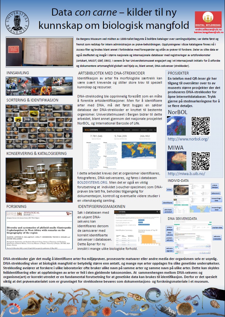
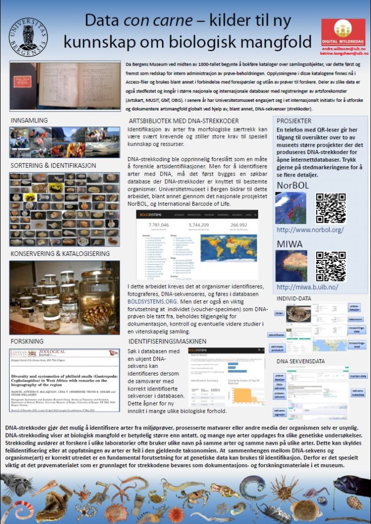
We brought with us a poster titled “Data con carne – sources of new knowledge on biodiversity” (in Norwegian), where we presented how our barcoding efforts on both African and Norwegian material are parts of a global undertaking of building a “library of life”, and how using huge databases such as BOLD can help us gain better understanding of biodiversity – and on where to focus our efforts in unraveling the taxonomy.

2017_digital-day

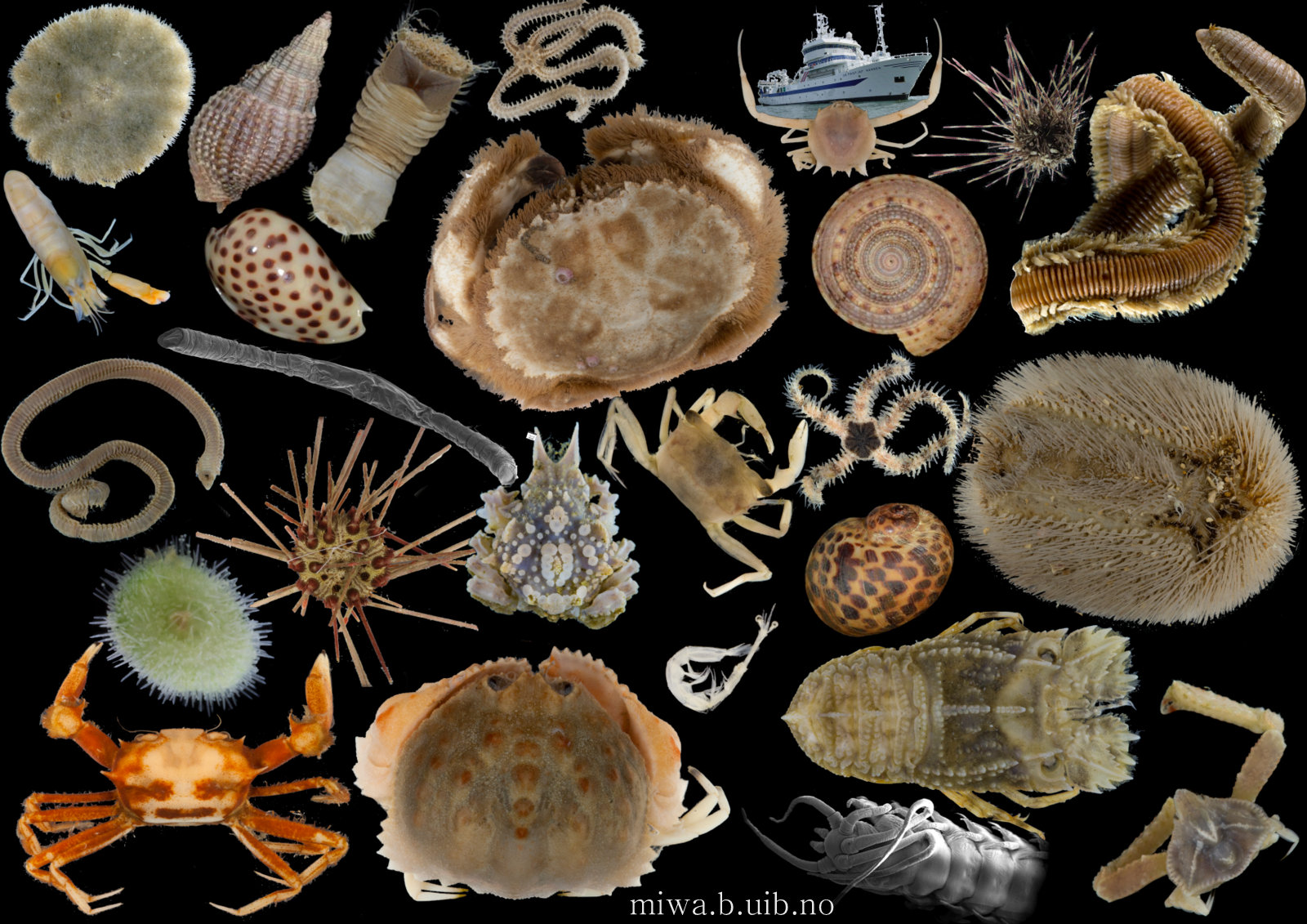
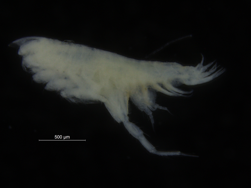
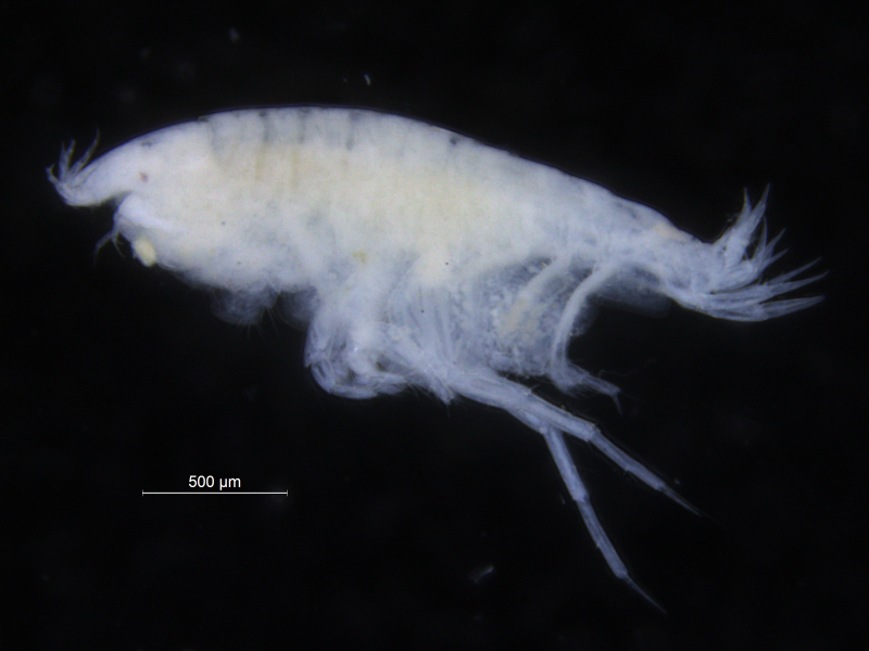
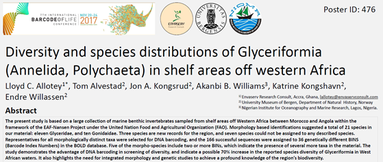
There is a very real challenge connected with estimating biodiversity when many of the species are still undescribed, as is the case with many invertebrates, especially the more obscure and diminutive groups.  In such cases, barcoding can serve as a tool in screening for biodiversity, and aid the taxonomists in identifying areas where the taxonomic resolution is poor.

There is a global effort underway to establish a library of short,  species specific genetic sequences. These standardized genetic sequences (“barcodes”) consists of a segment of approximately 650 base pairs of the mitochondrial gene cytochrome oxidase c subunit 1 (COI). You can read more about DNA barcoding on WIKIPEDIA.

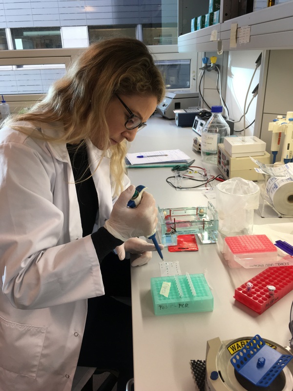
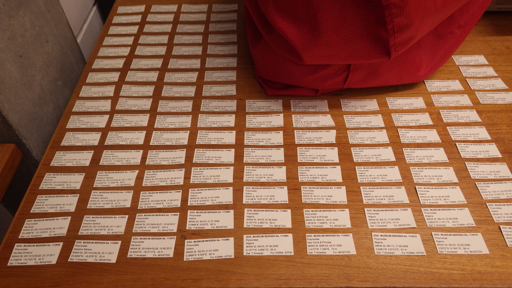
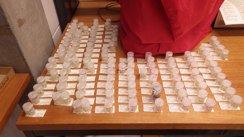
For MIWA, we have submitted over 2700 tissue samples from over 600 morphospecies for DNA barcode sequencing through the BOLD-pipeline, where the lab work is done in Guelph, Canada, and the data is uploaded to the Barcode of Life Data Systems (BOLD Systems) .

Out of these, 1450 sequences have been obtained (54% sequencing success rate), and these cluster into close to 550 BINs (OTUs) – suggesting that the diversity may be (much!) higher than what our preliminary investigations reveal. This is not so surprising, considering how hugely diverse and little studied the invertebrate fauna of the GCLME and the CCLME is.

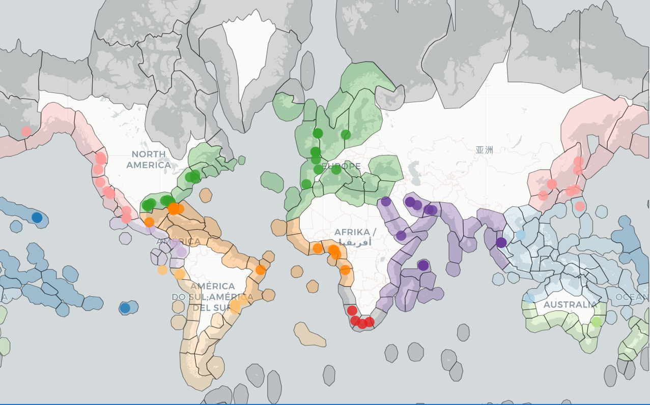
Below is an interactive map of the submitted samples, you can click on the stations to see the specimens that have been sent from that location, and whether or not they have gotten a barcode. You can also follow this link to find the map

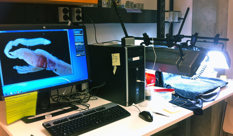
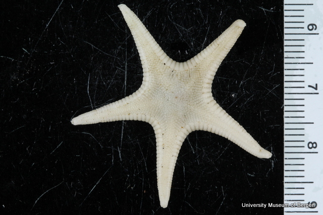
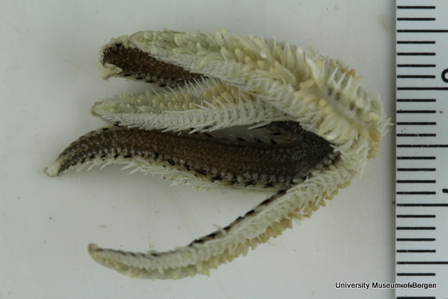
What we find is that it is crucial to be able to go back and reexamine the material post-barcoding, and that is where the “con carne” part of our poster came from;

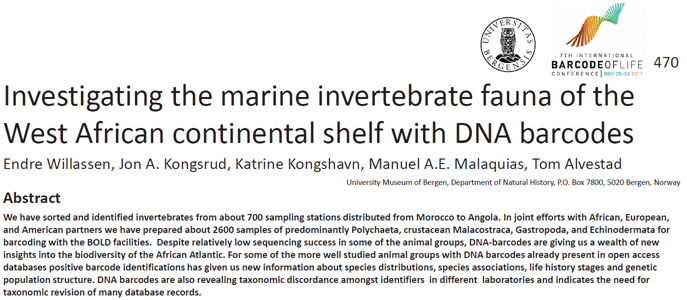
Our barcoding revealed several examples of mismatching taxonomic identifications amongst researchers in different labs and institutions in different countries. This illustrates a major challenge, as it has been shown that when benthic fauna is used in standardized methods for quality assessment and monitoring, mismatching identifications produced by different identifiers can have strong effects on indexing and valuation of ecological conditions in the same habitat.

For DNA barcoding to be useful, it is absolutely neccessary that the correlation between species name, specimen, and barcode is correct (or at least as close to it as we can get, if the species is undescribed).

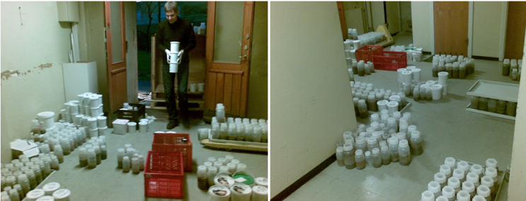
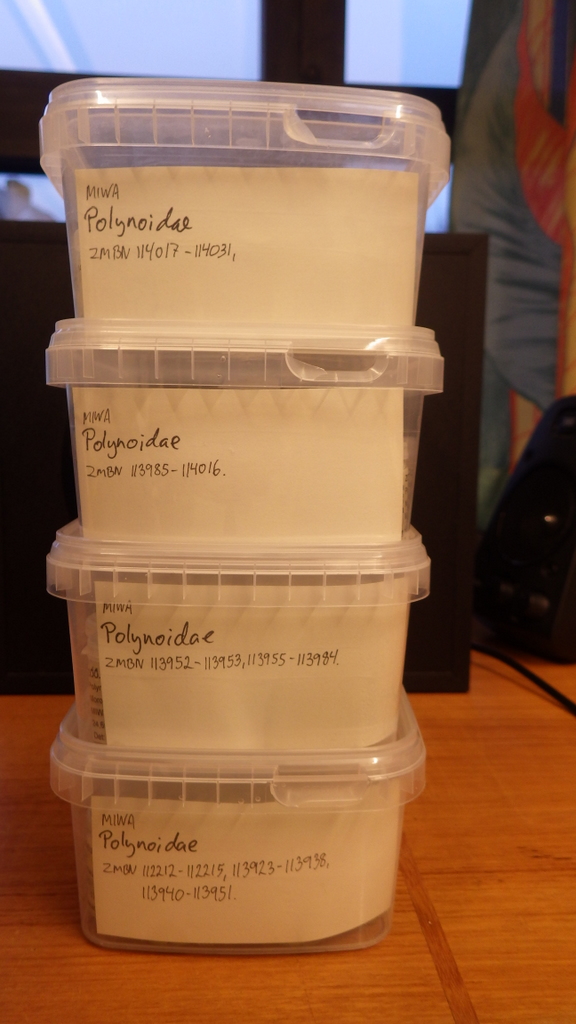
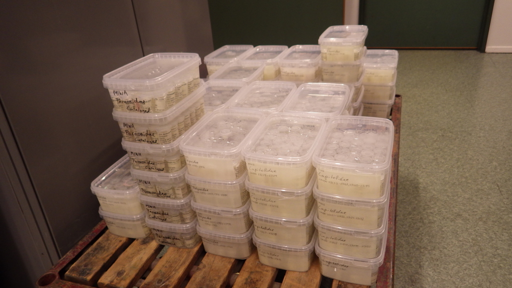
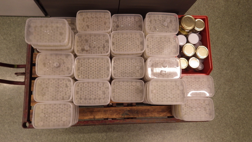
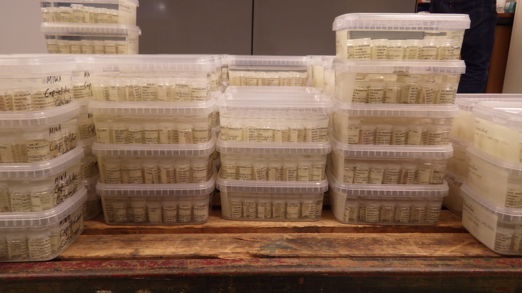
We must first build this reference library before barcoding can be reliably used for identification of unknown organisms. Hence it is imperative that the voucher specimens that correspond to the genetic barcodes are deposited in a museum where it will be preserved and made available for research.
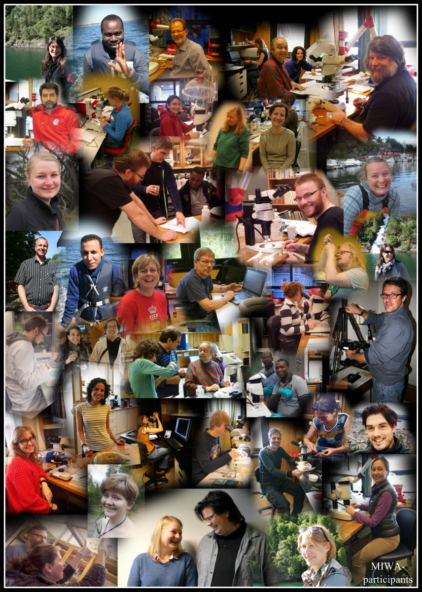
-Endre & Katrine

Indeed we did!

(get DNA from our faded stars, that is)
zmbn_115365_1

As explained in a previous post, we submitted tissues samples from the sea stars (class Asteroidea) that were suitable for DNA-analysis to the CCDB lab and the BOLD database. We just recieved the preliminary results, which are very promising!

The next step now will be to collaborate further with our asteroid taxonomist, and evaluate the genetic data combined with traditional morphology based taxonomy. Combining barcoding and morphology in this way helps us explore and understand the biodiversity better.

Stay tuned for updates!

Cataloging: the art of keeping track

Combining a multitude of guest researchers and our own efforts at working through the material, we’re producing thousands of new records to be cataloged into our database (we’ll get those listed here eventually, work is still ongoing at the moment).

Cataloging samples also means that the material needs to be physically updated;
New labels are printed for the samples,  and these must then be matched to their respective jars, inserted, and stored safely. It may sound pretty straight forward, but with this many samples, it is still a time consuming job.


Guest researchers: Polina

Polina, a master student jointly supervised by Dr. Nataliya Budaeva (UiB) and Dr. Alexander Tzetlin (Moscow State University) has spent a month in the invertebrate collections studying the bristle worms from the family Lumberineridae from the Western African Waters.

Lumbrinerid genera can be distinguished by the morphology of the jaw apparatus consisting of ventral fused mandibles and two rows of dorsal maxillary plates. Polina learned how to dissect the jaws and identified at least 11 genera of Lumbrinerids from the studied material. She is also planning to used microCT back at the Moscow University to study the morphology of the jaws in 3D. During her stay, Polina has studied the composition and morphology of chaetae, another character used in generic and species identification in Lumbrinerids, using SEM.

In addition, all studied specimens will be used in the molecular analysis to reconstruct the first phylogeny of the family Lumbrineridae based on genetic data. Please see the full description of the project: http://miwa.w.uib.no/allprojects/polychaeta-projects/

A – Ninoe sp. anterior part of body, ventral view; B – the same, close view of parapodia and chaetae; C – Gallardoneris sp., compound hooks; D – Scoletoma sp., anterior part of body.

Scanning Electron Microscope (SEM) images of Lumbrinerids: A – Ninoe sp. anterior part of body, ventral view; B – the same, close view of parapodia and chaetae; C – Gallardoneris sp., compound hooks; D – Scoletoma sp., anterior part of body.